Tissue Homogenization and Protein Extraction for Olink and SomaScan Proteomics
The last decade has witnessed significant growth of proteomics and its move toward population-scale and longitudinal studies, Olink and Somascan deliver broad proteome coverage with reproducibility and scalability and are becoming increasingly central to translational research and precision medicine. However, these high-plex technologies can only be as informative as the quality of protein sample presented for analysis. When it comes to sample matrices such as, tissues, FFPE , cells, organoids, and other complex biological matrices, researchers can face significant challenges in obtaining a consistent and representative protein sample.
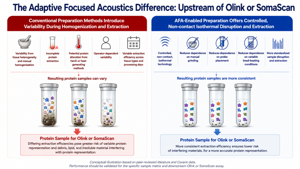
Tissue homogenization and protein extraction from tissue must account for differences in extracellular matrix, fibrosis, fat content, cellularity, and mechanical strength. These characteristics can affect the quality and quantity of proteins that are released and the consistency with which they are recovered across all sample types. Manual homogenization and other variable preparation steps can introduce additional differences between operators, specimens, and processing days.
For affinity-based proteomics, these effects have consequences beyond total protein yield. Proteins that are incompletely extracted, or whose assay-recognizable features are altered during preparation, may change the composition of the sample ultimately presented to Olink or SomaScan. The challenge is therefore to achieve reproducible extraction while maintaining compatibility with downstream protein detection.
Why tissue homogenization matters in high-plex proteomics
Tissue presents substantial structural complexity. Extracellular matrix composition, fibrosis, fat content, cellularity, and mechanical strength can all vary among tissue types and individual specimens.
These complexities and their inherent variability can influence the efficiency with which proteins are released during tissue homogenization and protein extraction. Manual grinding, probe-based disruption, bead beating, and other preparation approaches can also introduce variability between operators, samples, and processing days.
As a result, samples processed differently may produce proteins that differ not only in concentration, but also exhibit structural variability.
This makes a differente for high-plex proteomics studies, where structural and concentration differences may get interpreted as as issues pertaining to underlying biology.
Protein extraction from tissue is about more than total protein yield
A successful protein extraction from tissue or fresh frozen tissue samples should not be evaluated solely by how much total protein is recovered.
Consider two tissue lysates normalized to the same total protein concentration. One could contain fewer membrane-associated, extracellular, nuclear, or tightly bound proteins than the other because those proteins were not released with the same efficiency during extraction.
Protein normalization can correct differences in bulk concentration or assay loading. It cannot recover proteins that were never released from the tissue, correct selective loss of difficult-to-extract protein classes, or reverse matrix-specific extraction bias.
For this reason, standardization of tissue homogenization and protein extraction is important before protein quantification and normalization.
The sample preparation challenge for Olink and SomaScan
Olink and SomaScan use different approaches to protein detection. Olink uses pairs of protein-specific antibodies coupled to DNA oligonucleotides for proximity-based detection, whereas SomaScan uses engineered SOMAmer (modified DNA aptamer) reagents that bind directly to target proteins with high affinity and specificity.
This means protein extraction during the upstream sample preparation step must accomplish two objectives: effectively release proteins from the biological matrix, and preservethe features required for downstream recognition.
Aggressive or heat-generating preparation can introduce unnecessary thermal or mechanical stress. Probe-based approaches (in addition to contamination issues) may also create localized heating or excessive shearing that can potentially affect protein conformation or binding sites.
The objective of sample preparation, therefore, is not simply to produce more protein. It is to create a reliable, consistent and biologically representative protein sample for affinity-based measurement.
Using AFA® technology for controlled tissue homogenization and protein extraction
Covaris Adaptive Focused Acoustics® (AFA®) technology enables controlled, non-contact, isothermal disruption and extraction of complex biological matrices.
AFA-enabled workflows reduce dependence on variables associated with manual grinding, probe placement, or variable bead-beating conditions, standardizing sample disruption and protein extraction across a study.
This becomes particularly relevant when tissue characteristics differ substantially. Fibrotic, fatty, muscular, or otherwise mechanically difficult tissues can present different extraction challenges than softer or less structurally complex specimens.
A standardized disruption approach can help researchers address this matrix-dependent variability while using a buffer selected for compatibility with the downstream affinity assay. Researchers can explore additional approaches for reproducible protein extraction across sample types through Covaris proteomics sample preparation.
Prioritizing Sequencing Efficiency to Maximize Research Outcomes
Sequencing efficiency directly impacts research outcomes. Non-informative reads, uneven coverage, and rRNA impact research ROI by carrying overinflated sequencing depth requirements and increasing cost/usable transcript.
Optimized approaches focus on maximizing total amount of informative reads, preserving library complexity, and minimizing need for resequencing. Less technical noise means more usable data/run, immensely valuable for high-throughput projects and multi-sample cohorts. For many teams, that means prioritizing a streamlined RNA-seq workflow consisting of strong rRNA depletion, step-reduced handling, and reproducibility across variable inputs.
Extending high-plex proteomics to challenging tissue types
The importance of reproducible sample preparation becomes especially apparent as Olink and SomaScan workflows expand into more complex biological matrices
Fresh and frozen tissue
Fresh and frozen tissue requires reproducible tissue homogenization, protein extraction, and clarification. Variation in tissue composition and mechanical properties can make manual preparation difficult to reproduce across larger sample sets.
A controlled workflow can help standardize these preparation steps before protein quantification, normalization, and high-plex measurement. This becomes particularly relevant for limited sample quantities. Covaris has evaluated low-input fresh-frozen tissue proteomics using AFA-enabled processing for scalable MS-based proteomics. These data illustrate the broader sample preparation challenge of achieving reproducible homogenization and extraction from small quantities of tissue.
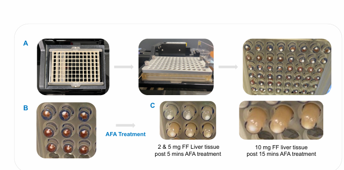
Figure 2. AFA-enabled homogenization of fresh-frozen tissue across a range of sample inputs. Fresh-frozen liver tissue at 2, 5, and 10 mg inputs before and after AFA processing using the Covaris R230 Focused-ultrasonicator. Consistent tissue homogenization was demonstrated across the tested input range. Data presented at HUPO World Congress 2025.
Tumor biopsies and heterogeneous tissues
Tumor biopsies may be small, heterogeneous, and high-value specimens. Variability in cellularity, extracellular matrix, fibrosis, or tissue composition can make consistent processing particularly important when researchers need to compare samples across a study.
Mechanically difficult tissues, including fibrotic, fatty, and muscular specimens, can present an additional risk of matrix-dependent under-extraction. For these samples, consistent tissue homogenization can help reduce the influence of sample preparation on the composition of the resulting protein extract.
FFPE tissue and FFPE proteomics
Formalin-fixed, paraffin-embedded tissue represents a particularly differentiated opportunity for high-plex proteomics because archived specimens can provide access to extensively characterized clinical sample collections.
At the same time, FFPE protein extraction presents distinct challenges. Crosslinking, dehydration, and paraffin embedding can complicate the recovery of proteins from archived specimens. Covaris FFPE extraction workflows use AFA technology for protein extraction from FFPE tissue, including active paraffin removal and tissue rehydration prior to protein purification.
The challenge can become even more pronounced when researchers have limited FFPE material available. Covaris has investigated proteomics sample preparation from small FFPE inputs, including workflows beginning with a single 10 µm FFPE scroll and AFA-assisted low-input FFPE proteomics workflows developed for MS-based analysis of limited FFPE samples.
These MS-based studies demonstrate approaches to the sample preparation challenges associated with limited FFPE material. For Olink and SomaScan applications, however, FFPE proteomics workflows require platform-specific validation of protein recovery and preservation of the binding features needed for affinity-based detection.
Beyond tissue: cell lysis for protein extraction from cells and organoids
Many of the same pre-analytical principles extend to cells and organoids.
Cell lysis for protein extraction can vary with cell type, cell number, operator, batch, and preparation conditions. As studies increase in size, seemingly small differences in manual processing can become important sources of technical variability.
A standardized approach to cell lysis can help create more consistent lysates across samples and provide a path from small feasibility studies toward larger, plate-based workflows.
Sample input is also an important consideration. One of the AFA-assisted workflow was evaluated for proteomics analysis from limited numbers of cells, including samples containing 1,000 and 10,000 cells. These data were generated using MS-based proteomics, but they demonstrate the application of controlled sample preparation to limited cellular inputs.
This makes upstream standardization relevant not only to conventional tissue proteomics, but also to studies using cultured cells, organoids, and other complex cellular models.
Standardization needs to happen before normalization
High-plex proteomics studies often depend on normalization to improve comparability among samples. But normalization and standardized sample preparation solve different problems.
Normalization can address concentration and loading differences. It cannot correct compositional bias introduced when proteins are extracted inconsistently from the original biological matrix.
Controlling tissue homogenization and protein extraction from tissue therefore addresses variability at the stage where compositional differences can be introduced.
The distinction becomes increasingly important in translational and cohort studies, where preparation methods must perform consistently across larger numbers of specimens, operators, processing days, and potentially, laboratories.
Scaling tissue homogenization and protein extraction for larger proteomics studies
A tissue homogenization workflow that works for a handful of samples may become difficult to execute consistently across hundreds or thousands.
Manual processing can introduce operator dependence and complicate transfer between laboratories. Increasing sample numbers also creates practical requirements for reduced hands-on processing, plate-based workflows, and automation compatibility.
AFA-enabled sample preparation on the Covaris R230 Focused-ultrasonicator provides a path from feasibility studies toward standardized, automation-compatible workflows for complex biological samples. Covaris has developed proteomics sample preparation workflows spanning fresh tissue, FFPE tissue, and cells, including approaches designed for low sample inputs and plate-based processing.
For researchers evaluating a sample preparation workflow for Olink or SomaScan, useful validation criteria can include:
- Technical reproducibility
- Protein detectability
- Biological fidelity
- Protein recovery
- Binding-site preservation
- Matrix robustness
- Workflow performance
- Cross-site transferability
Better sample preparation supports greater confidence in the biology
Olink and SomaScan provide powerful tools for high-plex protein detection. Downstream analytical capability, however, cannot compensate for proteins that were not reproducibly extracted from the original sample.
For tissue, FFPE, cells, and other complex matrices, the objective is broader than maximizing total protein yield. Researchers need tissue homogenization and protein extraction workflows that are reproducible, representative, scalable, and compatible with affinity-based detection.
By addressing variability before protein quantification and normalization, Covaris AFA-enabled sample preparation helps researchers create more consistent inputs for Olink and SomaScan. This increases confidence that measured differences reflect the biology of the original sample, rather than variability introduced during preparation.
Learn more about Covaris proteomics sample preparation solutions for reproducible protein extraction and processing across complex biological sample types.
Speak to a Covaris proteomics sample prep expert
Olink® and SomaScanTM are registered trademarks of their respective owners. Covaris is not affiliated with or sponsored by Olink or SomaLogic.