AFA Assisted Workflow for High Throughput Mass Spectrometry-Based Proteomic Analysis of FFPE Samples
At ASMS 2025, Dr. Anu Jain, a postdoctoral fellow at the Mayo Clinic, shared how her team is simplifying proteomic analysis of formalin-fixed paraffin-embedded (FFPE) samples using sample preparation workflows enabled by Covaris’ Adaptive Focused Acoustics (AFA) technology. With recent advancements in mass spectrometry, it has become feasible to explore the cellular proteome at a low number of cells or even single cells. Dr. Jain and her team’s objective was to develop a robust and reliable sample preparation workflow for analyzing the proteome obtained from a limited number of cells, starting with FFPE tissue. By rethinking traditional workflows, they’re achieving faster digestion, better protein yields, and reliable results—even from ultra-low input samples. In this blog, we explore how AFA is helping researchers maximize the benefits of FFPE samples, without the usual trade-offs.
Introduction
Formalin-fixed paraffin-embedded (FFPE) tissue samples have long been considered a rich resource for proteomics research because of their abundance, long-term stability, and valuable clinical annotations. However, working with FFPE samples can be challenging, as traditional methods are slow, labor-intensive, and difficult to scale up, especially for large batches comprising small input amounts. During Dr. Jain’s ASMS presentation, she demonstrated several unique and robust AFA-enabled workflows that address these traditional sample preparation challenges with a more efficient and reproducible approach.
Rethinking Sample Prep with AFA Technology
To improve throughput while ensuring consistent data quality, Dr. Jain adopted the Covaris R230 focused-ultrasonicator, which uses adaptive focused acoustics (AFA) to streamline and improve sample processing
Two key advantages stood out:
- Format Flexibility: R230’s compatibility with 8-strip tubes, 96-well or 384-well plates makes it suitable for small-scale studies for method development and also addresses high-throughput sample pipelines.
- Temperature Control: Covaris’s water bath ensures good thermal control for all sample types and throughout the sample preparation workflow; a critical factor in preserving protein integrity, enzyme activity, and ultimately ensuring reproducibility.
Their Leica laser capture microdissection (LCM) system was also adapted to directly collect FFPE tissue samples into Covaris AFA-compatible plates, eliminating the need for transfer steps and reducing the chances of losing precious samples.
Comparing Workflows: Standard vs. AFA-Assisted
To test the impact of AFA, Dr. Jain designed a comparative study using 48 LCM samples from a single FFPE block, and divided the total number of samples into three different methods (Figure 1):
- Standard: Collected in microfuge tubes with overnight digestion
- AFA Method #1: Overnight digestion with AFA applied pre- and post-decrosslinking
- AFA Method #2: One-hour digestion with AFA applied during digestion

Figure 1. Evaluation of AFA-assisted method.
The results spoke for themselves (Figure 2):
- The standard method identified over 1,000 proteins.
- Both AFA-assisted methods pushed that number to nearly 1,500 proteins.
- Missed cleavage rates remained consistent across all groups, with high reproducibility across 16 replicates.
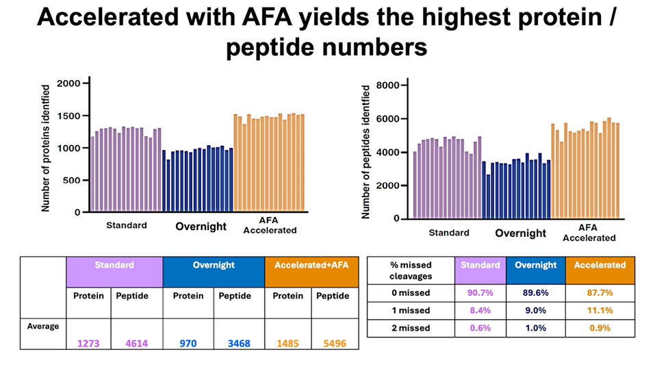
Figure 2. Accelerated with AFA yields the highest protein/peptide numbers.
The takeaway: AFA-assisted digestion not only saves time; it delivers better data (higher protein coverage, better peptide depth) without sacrificing quality.
Next Step—Going Smaller
Dr. Jain went beyond improving workflow speed. She also evaluated the sensitivity of her mass spectrometer and the ability of the AFA-enabled workflow to obtain high-quality data starting with highly limited input samples. From tissue areas as small as 0.5 mm², Dr. Jain identified nearly 1,000 proteins. Increasing the area to just 1 mm² yielded close to 2,000 proteins!
Building on these results, the team transitioned to a 384-well plate format and shifted from small tissue sections to precise cell counts. They tested AFA-assisted workflows (including digestion) on 10, 50, and 100 colon tissue cells (Figure 3).

Figure 3. Analysis of 10, 50, and 100 cells from FFPE tissue samples.
- From 100 cells, they identified ~4,000 proteins and ~20,000 peptides (Figure 4).
- From 10 cells, they still identified ~2,000 proteins and ~10,000 peptides (Figure 4).
- For each of the peptides observed, the peptide lengths were consistent, mostly between 7–12 amino acids (Figure 5).
- Most importantly, the data was reproducible across replicates, even at such ultra-low input levels.
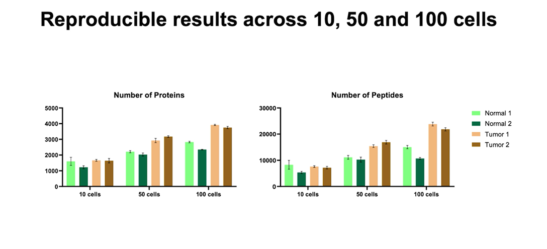
Figure 4. Reproducible results across 10, 50 and 100 cells.

Figure 5. Analysis of 10, 50, and 100 cells from FFPE tissue samples.
While reproducible and reliable single-cell workflows are still being optimized, early data shows real promise for applying AFA in ultra-low input proteomics.
Streamlining Sample Handling with a Single-Plate Workflow
One major strength of the AFA-enabled workflow is its ability to work within a single tube/plate, minimizing sample handling. Cells can be collected, processed, and injected into the LC-MS system from the same 96- or 384-well plate. This ease-of-sample processing eliminates the need for transfers between vessels, reducing sample loss—often the biggest challenge in low-input sample workflows.
Dr. Jain emphasized that this streamlined, single-plate format not only reduces variability but also lends itself well to automation, which is key for scaling up.
Scaling Up: From Sections to Punches
The Covaris AFA method isn’t just effective for micro-dissected sections. Dr. Jain also applied it to tissue punches, which are small cylindrical samples taken from FFPE blocks. Using AFA-compatible bead snap cap tubes with a single ceramic homogenization bead, they processed 1×1 mm punches from human cerebral cortex (Figure 6).
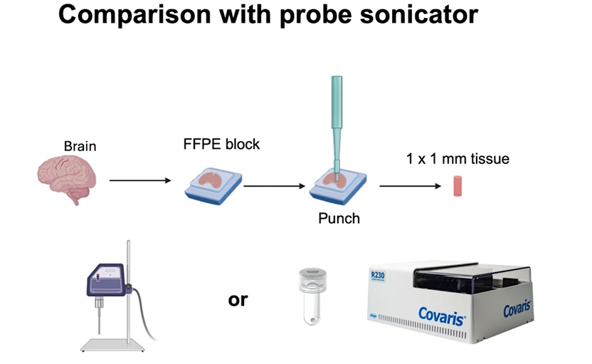
Figure 6. Comparison with probe sonicator.
The results held up (Figure 7):
- AFA-based homogenization matched the performance of traditional probe sonication.
- Unlike probe sonication, AFA offers consistent, temperature-controlled, and automatable processing. This specific feature ensures sample integrity and assay reliability.
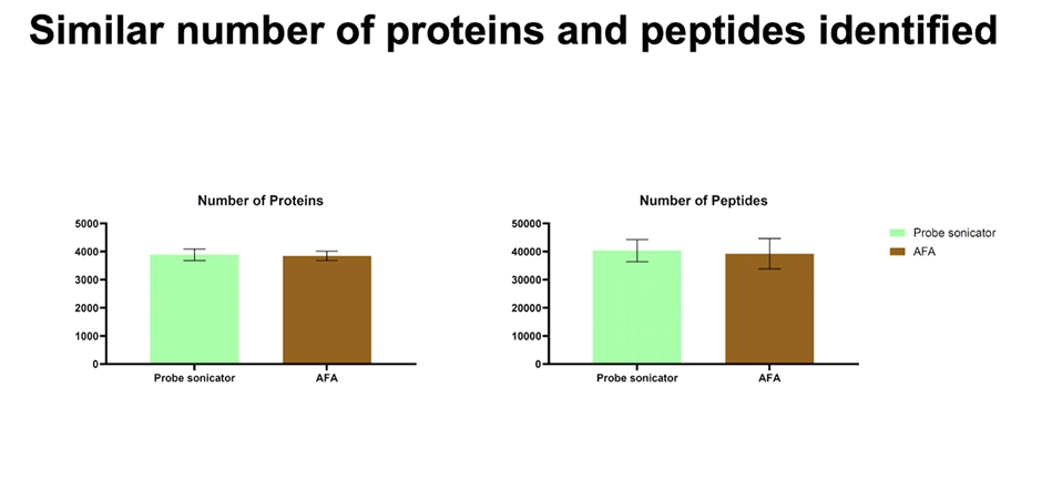
Figure 7. Similar number of proteins and peptides identified.
Peptide length distributions and missed cleavage rates were comparable between the two methods, confirming AFA’s versatility across tissue formats.
AFA Delivers Speed, Sensitivity, and Scalability
Dr. Jain concluded by saying: “We are able to get reproducible and sensitive data using AFA-assisted methods for FFPE analysis. These workflows offer high throughput potential and are compatible with automation.”
- One-hour digestion workflows that outperform traditional overnight protocols
- Direct processing in 96- or 384-well plates, eliminating sample loss
- Reproducible Low-input analysis with high sensitivity and reproducibility
- Automated homogenization for both sections and punches, reducing variability
The combination of Covaris hardware and AFA-enabled protocols is helping proteomics labs push the boundaries of what’s possible with FFPE samples, whether in large cohort studies or ultra-sensitive single-cell work.
Click here to watch Dr. Jain’s full presentation: https://youtu.be/3x4lX5ydoXQ?si=3i8ibtfmP4oJsgJ3 [/vc_column_text][/vc_column][/vc_row]