Simplified Homogenization Workflow to Maximize Protein Extraction and Reduce Variance with AFA Technology
Proteomic analysis plays a critical role in drug development, but the quality of that data hinges on how well tissue samples are prepared. At ASMS 2025, Juan Wang, PhD, Senior Scientist at AstraZeneca, presented two optimized workflows that use Adaptive Focused Acoustics® (AFA®) technology, with and without dry pulverization, to address the long-standing challenges of bead beating. Her team’s approach not only improved protein yields and reduced variability across tissue types, but also cut hands-on time by nearly 45%, offering a more consistent and scalable way to prepare tissue samples for mass spectrometry.
Introduction
In proteomics research, what happens before mass spectrometry can make or break results. That’s especially true when you’re working with complex biological samples, where the goal is to efficiently maximize total yield of high-quality protein from a wide variety of tissue types without introducing variability and ensuring impactful data leading to confident results. This is where many labs struggle. Bead beating, the standard technique for homogenizing tissue, is far from perfect. It’s hard to standardize, it generates heat that can compromise samples (especially proteins), and requires constant hands-on attention.
Recognizing this, Dr. Wang and her team at AstraZeneca set out to build a better solution, hoping to mitigate the challenges of bead beating. At ASMS 2025, she presented two streamlined workflows that used Covaris AFA® technology to automate tissue homogenization (with or without cryoPREP® dry pulverization based on the amount of tissue input). The goal was straightforward: improve consistency, increase protein yields, and make the process easier to scale.
Two Workflows Built Around Real-World Needs
To accommodate a range of experimental designs, the team developed two distinct workflows (Figures 1-3):
Workflow 1: High-Precision, Low-Throughput (24 samples)
- Ideal for small sample batches requiring ultra-consistent results
- Includes cryoPREP dry pulverization + AFA homogenization
- Tissue amount: ~50 mg or higher
- Applications: Comparative studies across different tissue types or conditions, Targeted analysis

Figure 1. Current sample preparation workflow (1) – different tissues: liver, brain, kidney, heart, pancreas, intestine, spleen.
This workflow consistently produced high amounts of protein and low variance across liver, kidney, brain, spleen, heart, and pancreas samples. It’s the go-to option when data quality is paramount when working with larger tissue input amounts.
Workflow 2: High-Throughput (96 samples)
- Ideal for large-scale studies and screening projects
- Excludes dry pulverization to streamline processing
- Tissue amount: 1–10 mg (using biopsy punches)
- Applications: Time-sensitive experiments, large cohorts

Figure 2. New sample preparation workflow (2) is developed.

Figure 3. Comparison of workflows 1 and 2.
Despite skipping the cryoPREP step, Workflow 2 still delivered excellent protein extraction, especially when combined with biopsy punches for consistent tissue input, which eliminated the need for weighing tissue manually. With diameters of 1.0–1.5 mm, the researchers could pull 2–9 mg of tissue depending on thickness, making plate setup faster and more uniform across wells. Even in this accelerated format, the CVs remained under 15%, demonstrating the robustness of the AFA method.
Optimizing AFA® Parameters for Maximum Protein Recovery
To get the most out of AFA, the team optimized three key parameters (Figure 4):
- Peak Incident Power (PIP): The maximum acoustic energy amplitude applied to the sample.
- Duty Factor: The percentage of time the acoustic energy is actively delivered.
- Cycles Per Burst (CPB): The number of acoustic pulses delivered per active phase.
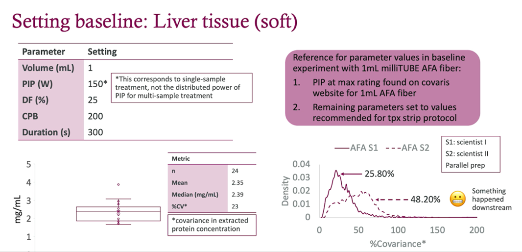
Figure 4. Setting baseline: Liver tissue (soft).
Through iterative testing, the team of researchers identified a powerful parameter set (PIP 450, Duty Factor 50%, CPB 1000) that delivered improved results. Compared to earlier protocols, protein yield increased from ~2–3 mg/mL to 6–8 mg/mL, and coefficients of variation (CVs) dropped to below 10% in soft tissues and under 20% in harder tissues. This precision directly translated into improved mass spectrometry data quality (Figures 5-7).
Results That Hold Up Across Sample Types
Across both workflows, the Covaris-enabled protocols consistently delivered:
- High protein concentration: 4–8 mg/mL across tissue types
- Low variance: %CVs as low as 8% for soft tissues (e.g., spleen, intestine), and below 20% for harder tissues (e.g., heart, liver)
- Better protein ID counts: Compared to bead beating or early-stage AFA parameters
The optimized workflows were demonstrated to be broadly applicable to tissues of varying types. The new high-throughput workflow decreased the fraction of time spent on active engagement by approximately 45%, achieving the target goal.
These improvements had direct impacts on downstream proteomics, where reproducible extraction leads to more reliable quantification and deeper insights.

Figure 5. Workflow 1: extracted protein concentrations and variance with liver tissues.
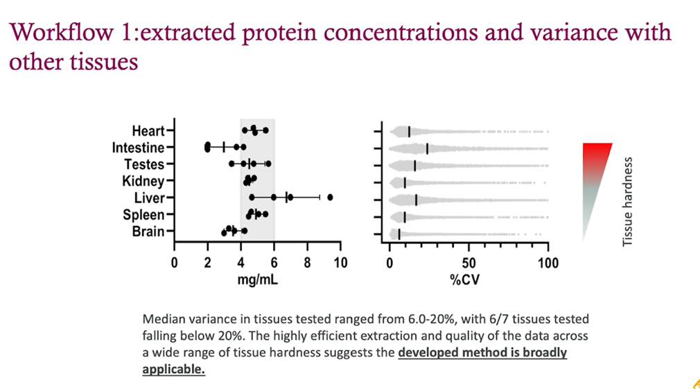
Figure 6. Workflow 1: extracted protein concentrations and variance with other tissues.

Figure 7. Workflow 2: extracted protein concentrations and variance.
Conclusion
Dr. Wang’s team showed what’s possible when sample prep gets the same attention as the rest of the workflow. By moving beyond bead beating and adopting more controlled, automated approaches, labs can get more consistent protein extraction and higher-quality data, no matter the tissue type.
Whether you’re running a small exploratory study or processing hundreds of samples, Covaris AFA® technology provides a streamlined, high-performance foundation for global proteomics.
Watch Dr. Wang’s full presentation:
[/vc_column_text][/vc_column][/vc_row]