At ASMS 2023, Benjamin Madden, PhD, Staff Scientist at the Mayo Clinic Proteomics Core, presented results of a simplified laser capture microdissection (LCM) FFPE tissue processing workflow developed for proteomics research.
The work, conducted within the Mayo Clinic Proteomics Core, directed by Dr. Akhilesh Pandey, incorporated Adaptive Focused Acoustics® (AFA®) technology-assisted digestion using Covaris’ R230 Focused-ultrasonicator.
The use of a plate-based capture approach with AFA Technology reduced processing time without sacrificing protein and peptide identification numbers or reproducibility, enabling consistent detection of previously reported, disease-associated antigens in kidney tissue samples.
Introduction
High-quality protein recovery workflows from formalin-fixed paraffin-embedded (FFPE) tissue samples remain key to advancing proteomics research. Applying laser capture microdissection (LCM) to FFPE tissue isolates specific histological regions for mass spectrometry (MS)-based proteomic analysis in tumors. However, LCM presents challenges of laborious, time-consuming sample processing and low protein yields.
At the Mayo Clinic, Dr. Pandey’s team developed a faster and more robust automation-friendly workflow for LCM-FFPE tissue processing using AFA technology. Read on to learn how they integrated the Covaris R230 Focused-ultrasonicator to increase throughput and lower sample handling times without sacrificing performance.
Answering the Call for a Streamlined, Automation-Friendly LCM-FFPE Processing Workflow
Faced with increasing demand for LCM-FFPE processing, Dr. Pandey’s group needed to scale up their labor-intensive, standard tube-based workflow, which involved multiple manual steps and was not practical for processing in 96-well plates.
As project requests grew, the team outlined several primary goals for scaling up their workflow:
- Decrease processing time
- Consolidate collection and processing steps into the same device
- Achieve LCM capture directly into a 96-well plate
- Build in automation options
They were interested in leveraging the Covaris R230 Focused-ultrasonicator because it enabled exploration of both AFA technology and plate-based processing together. The R230’s design was especially amenable to their LCM method, which used the Leica LMD6 and a holder designed specifically for the Covaris 96 AFA-TUBE TPX plate (Figure 1).

Figure 1. Combining the R230 Focused-ultrasonicator’s plate-based format with Leica Microsystems’ LMD6 laser microdissection microscope for enabling sample capture directly into a 96-well plate.
Evaluating the Performance of AFA-Accelerated FFPE Protein Extraction in a 96-Well Format Compared to Standard Workflow
After selecting and integrating their tools, the team evaluated different processing options to determine how they could level up their existing proteomics workflow. In the study, 20 mm2 of normal tubulointerstitium FFPE kidney tissue was deparaffinized and microdissected and processed under five conditions (Figure 2):
- Standard workflow: Heat-induced retrieval and reduction, alkylation, and overnight trypsin digestion
- AFA overnight trypsin: Extraction using the R230 followed by overnight trypsin digestion
- AFA accelerated trypsin: Extraction using the R230 and simultaneous trypsin digestion using AFA (x 60 minutes) with R230
- SP3 + overnight trypsin: Adding a magnetic bead-based digestion method to the standard workflow
- SP3 + AFA accelerated trypsin: Adding a magnetic bead-based digestion method to the AFA accelerated method
Four replicates were processed under each condition. In this phase, all samples were first collected in tubes and then manually transferred to the 96-well plates for AFA processing. After digestion, the samples were cleaned and analyzed by mass spectrometry. Protein and peptide identification numbers, reproducibility, and processing time were compared across all workflows.

Figure 2. Comparison of sample preparation workflows for LCM-FFPE tissue processing.
Protein and Peptide Numbers in AFA-Accelerated Workflows Were Comparable to Standard Workflow
Mass spectrometry results showed that both AFA-accelerated methods performed as well as the standard tube-based method in terms of protein and peptide identification numbers (Figure 3).
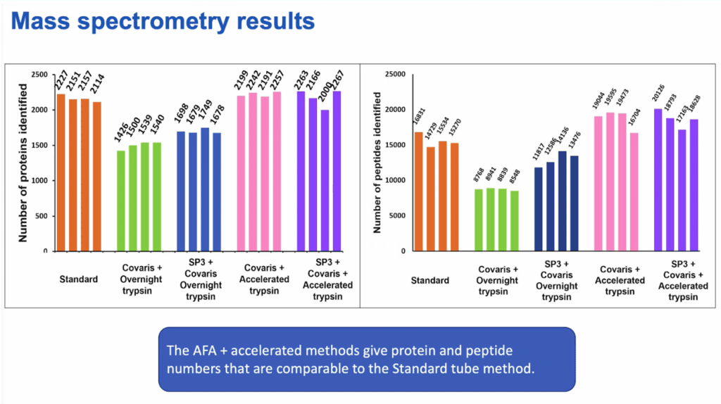
Figure 3. Both AFA-accelerated methods yielded protein and peptide numbers comparable to the standard method. Using SP3 did not add significant benefits.
These results showed that the researchers could save time by using faster accelerated digestion methods without sacrificing protein and peptide identification numbers.
AFA-Assisted Workflows Identified Most Protein and Peptide Numbers When Samples Were Collected Directly into 96-well Plates
Next, they tested whether collecting samples directly into the TPX Plate on the Leica® system affected protein and peptide identification numbers. Three conditions were tested using 16 replicates (Figure 4):
- Standard workflow (collecting into 0.5 mL microcentrifuge tubes)
- Collecting into the 96-well AFA-TUBE TPX Plate + AFA overnight trypsin
- Collecting into the 96-well AFA-TUBE TPX Plate + AFA-accelerated trypsin

Figure 4. Comparison of sample preparation workflows to test plate format.
MS results showed that when samples were collected directly into the TPX Plate, protein and peptide numbers were higher in the AFA-accelerated workflow compared to the standard workflow. Sample-to-sample variability was also lowest in the AFA-accelerated condition. Furthermore, the number of missed cleavages was very similar across all three workflows (Figure 5).

Figure 5. Mass spectrometry results showed that the AFA-accelerated workflow yielded a higher and more reproducible number of protein and peptide identifications.
Therefore, the team concluded that the AFA-accelerated, 96-well Plate format workflow enabled by the R230:
- Dramatically shortened processing time by 85% with no effect on protein and peptide numbers and little effect on number of missed cleavages
- Delivered consistent, high-throughput analysis with good sample-to-sample reproducibility
Simplifying to a Streamlined, All-in-One, Single-Well FFPE Processing Workflow
To explore whether a simple single-well workflow–where the sample is collected, processed, and injected from the same well–would be a viable option for scaling throughput, Dr. Pandey’s group executed a proof-of-concept experiment.
They collected 0.5 mm2 and 1.0 mm2 kidney glomeruli samples—tiny samples that reflected the distinct features of these structures and yielded fewer proteins and peptides. This workflow leveraged the Agilent Bravo liquid handling platform for reagent transfers, and manual handling for other transfers (Figure 6). The team was driven by the potential of processing 8–96 samples in just three hours end-to-end for their work that involved glomeruli-associated diseases.

Figure 6. All-in-one single-well FFPE processing workflow using the Leica LMD6, Agilent® Bravo, and Covaris R230.
Again, the results showed strong and consistent protein and peptide identification numbers from both 0.5 mm2 and 1.0 mm2 FFPE samples. Based on prior experience within the Mayo Clinic Proteomics Core, laser-capture microdissected FFPE tissue areas on the order of 1 mm² typically yield less than a microgram of total peptides for downstream proteomic analysis; the results obtained with this workflow indicate good analytical sensitivity. (Figure 7).

Figure 7. Mass spectrometry results for a three hour end-to-end single-well processing workflow.
Expanding the AFA-Accelerated Single-Well Workflow to Clinically-derived FFPE Research Samples
To test whether this single-well method could identify antigens in clinically-derived FFPE samples, the researchers drew from ongoing work on membranous nephropathy, a kidney disease characterized by the deposition of immune complexes across the glomerular basement membrane. They applied the newly developed single-well method to two relevant antigens they had previously identified: exostosin 1/exostosin 2 and NELL-1.
In a blind exploration, four FFPE tissue samples from each discovery cohort were analyzed using the single-well workflow. MS results showed that the single-well method identified the same antigens previously reported in each cohort. And again, the AFA-accelerated method results were comparable to the standard tube-based overnight digestion method and demonstrated no loss in sensitivity (Figure 8).
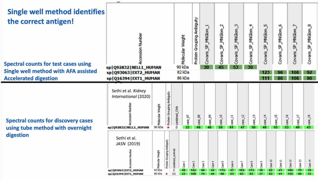
Figure 8. Spectral count results for each protein were comparable across the AFA-accelerated workflow and standard workflow.
AFA Combined with Single-Well Capture Offers Superior LCM-FFPE Performance Compared to Standard Workflow
Across these studies, Dr. Pandey’s team demonstrated that AFA-accelerated LCM-FFPE tissue protein extraction in a 96-well format produced:
- No sacrifice in protein and peptide numbers and sample-to-sample variability compared to a standard tube-based workflow
- Significant time savings with processing times of three hours or less
- Consistent results across replicates
- Automation-friendly integration
- Potential to identify antigens in FFPE research samples
Conclusion
Overall, results from the Mayo Clinic Proteomics Core pointed to the advantages of integrating the Covaris R230 Focused-ultrasonicator with the Leica LMD6 into an LCM-FFPE processing workflow, offering advantages in protein and peptide identification numbers and method simplicity, robustness, and reproducibility.
Ready to learn more about the Covaris R230 Focused-ultrasonicator and how to integrate AFA technology into your FFPE processing workflow? Contact one of our specialists.
Research Use Only (RUO) Notice:
The workflows, methods, and results described in this article are intended for research use only. Covaris technologies are not intended for diagnostic procedures or clinical decision-making. Any references to disease states or clinical samples are presented solely in the context of proteomics research and method evaluation.